
Overview
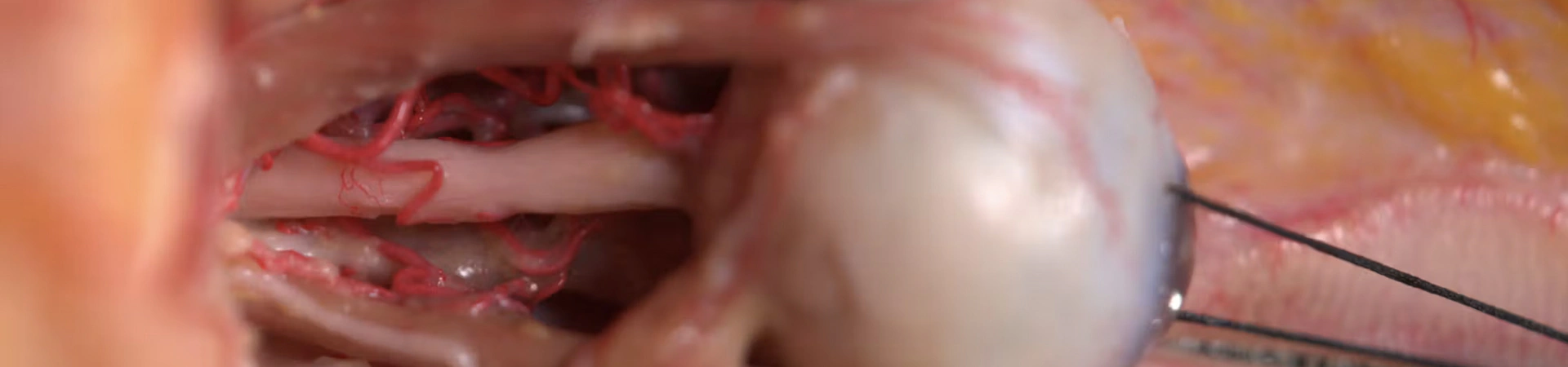
The periorbital region is directly connected to the temple and the forehead via direct fascial layers and via the arterial vascular network. In addition, it must provide a high degree of flexibility for the movement of the eyelids. All these factors together, predispose the periorbital region to be a challenging facial region to address with soft tissue fillers. Although no injection simulations are performed, this video is best suited for briefly freshen up on periorbital anatomy prior to clinical treatments.
Treatment type: Relevant for Soft Tissue Filler Injections
Facial region: Periorbital
Approach: Anatomic Review
Competency Level: Novice
Course duration: 27 minutes
Course type: Anatomic review on a pre-dissected body donor performed by Prof. Sebastian Cotofana
Learning Objectives
- Identify safe facial fascial planes to minimize injection risks.
- Explain the significance of key arterial pathways in the periorbital region.
- Identify and avoid high-risk zones during facial filler procedures.
- Differentiate facial planes to determine optimal injection sites of the periorbital region.
FAQs
Our online anatomy courses are designed for aesthetic practitioners, including MDs, RNs, PAs, and NPs, as well as medical students, residents, and any healthcare professionals.

Access the full
course library
Unlock the complete Cotofana Anatomy e-learning library with a monthly or annual membership.

Empower your
clinical team
Provide your organisation to the e-learning platform with a tailored Corporate Plan.
Buy this lesson
30-day access
Become a member
Get unlimited access