
Overview
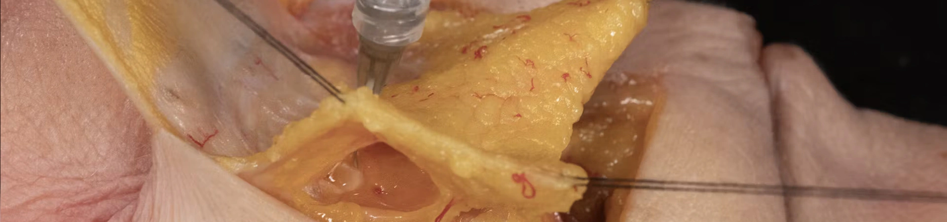
The palpebromalar groove divides the cheeks from the lower eyelid. The underlying anatomic structure that causes the formation of this groove is the orbicularis retaining ligament. Understanding that this grove is natural and normal to have, creates aesthetic difficulties when addressing this aesthetic concern. In this video, various techniques utilizing both needles and cannulas are demonstrated focusing on anatomy and on the clinical outcome.
Treatment type: Soft Tissue Fillers
Facial region: Periorbital
Approach: Cannula and Needle Injection Techniques
Competency Level: Competent
Course duration: 13 minutes
Course type: Injection simulation on a human body donor performed by Prof. Sebastian Cotofana
Learning Objectives
- Identify key anatomical landmarks relevant to lateral infraorbital anatomic region.
- Locate appropriate dermal access points and injection planes for infraorbital filler placement.
- Describe proper techniques for both needle and cannula advancement in the lateral infraorbital region.
- Confirm accurate product placement to ensure safety and effectiveness in treating lateral infraorbital hollows.
FAQs
Our online anatomy courses are designed for aesthetic practitioners, including MDs, RNs, PAs, and NPs, as well as medical students, residents, and any healthcare professionals.

Access the full
course library
Unlock the complete Cotofana Anatomy e-learning library with a monthly or annual membership.

Empower your
clinical team
Provide your organisation to the e-learning platform with a tailored Corporate Plan.
Buy this lesson
30-day access
Become a member
Get unlimited access